
X Rays Imaging For Body Scanning
X-ray imaging uses high-energy rays to create detailed pictures of the inside of the body, helping doctors diagnose and monitor various medical conditions.
What is X Rays
X-rays screening is a medical imaging technique.
- It uses X-rays, a form of electromagnetic radiation.
- X-rays pass through the body and create images on film or digital detectors.
- It's used to visualize internal structures like bones, organs, and tissues.
- Commonly used for diagnosing fractures, lung conditions, and dental issues.
- Low-dose X-rays are generally safe but involve some radiation exposure.
- Controlled use in healthcare provides valuable diagnostic information.
X Rays
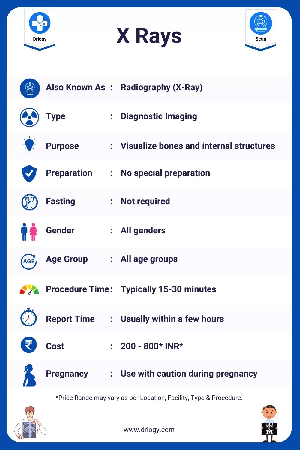
Here are the basic details for the X Rays.
| Also Known As | Radiography (X-Ray) |
| Type | Diagnostic Imaging |
| Purpose | Visualize bones and internal structures |
| Preparation | No special preparation |
| Fasting | Not required |
| Gender | All genders |
| Age Group | All age groups |
| Procedure Duration | Typically 15-30 minutes |
| Reporting Time | Usually within a few hours |
| Cost | 200 - 800* INR* |
| Pregnancy Consideration | Use with caution during pregnancy |
| Risks and Safety | Low radiation exposure, generally safe |
| Accessibility | Widely available in medical facilities |
*Price range may vary as per location, facility, type, and procedure.
What are the Purpose or Reasons for X Rays?
Here are common reasons for X Rays.
- Visualize and assess bone fractures and injuries
- Detect and monitor lung conditions and infections
- Diagnose dental and oral health issues
- Screen for and evaluate chest and abdominal conditions
- Guide the placement of medical devices and implants
- Monitor changes in the skeletal system over time
- Identify foreign objects or abnormalities in the body
Types of X Ray Scans
Here are the types of X Rays scan along with their primary use.
| X-Ray Scan Type | Organ/System | Primary Use |
|---|---|---|
| Chest X-Ray | Chest/lungs | Evaluate lung and heart health |
| Abdominal X-Ray | Abdomen | Assess abdominal organ function |
| Dental X-Rays | Teeth/jaws | Diagnose dental and oral issues |
| Skeletal X-Rays | Bones/joints | Detect fractures and bone issues |
| Spinal X-Ray | Spine | Evaluate spinal health and injury |
| Barium Swallow | Esophagus | Visualize swallowing mechanisms |
| Mammography | Breasts | Detect breast abnormalities |
| CT Scan (Computed Tomography) | Various | Visualize internal structures |
| MRI (Magnetic Resonance Imaging) | Various | Detailed imaging for diagnosis |
These X-ray scan types serve various diagnostic purposes across different organ systems and conditions.
Preparing for Your X Rays: Tips and Information
Here is the basic preparation before, during, and after X Rays for any patient.
Before the X-Ray:
- Consultation: Schedule the X-ray and discuss your medical history and any concerns with your healthcare provider.
- Clothing: Wear comfortable clothing without metal fasteners, zippers, or buttons in the area to be X-rayed.
- Jewelry and Accessories: Remove all jewelry and accessories in the area to be X-rayed.
- Pregnancy: If you are pregnant or suspect pregnancy, inform your healthcare provider, as special precautions may be necessary.
During the X-Ray:
- Positioning: You will be positioned by a radiologic technologist on the X-ray table. They may use protective lead aprons to shield areas not being examined.
- Communication: Follow the technologist's instructions for proper positioning and holding your breath when needed. Communicate any discomfort during the procedure.
- Remain Still: It's important to stay as still as possible to obtain clear X-ray images.
After the X-Ray:
- Recovery: There is typically no special recovery required, and you can usually resume normal activities immediately.
- Results: Your X-ray results will be reviewed by a radiologist, and a report will be sent to your healthcare provider.
- Follow-Up: Schedule a follow-up appointment with your healthcare provider to discuss the X-ray results and any further steps or treatments if needed.
Please note that X-ray procedures are generally straightforward and do not require extensive preparation or recovery. Specific instructions may vary based on the type of X-ray and the area being examined.
Who Performs a X Rays?
| Professional | Role |
|---|---|
| Radiologic Technologist | Takes X-ray images. |
| Radiologist | Interprets X-ray results, provides a report. |
| Radiology Nurse | Assists patients during X-ray procedures. |
X Rays Procedure
The procedure for X Rays typically follows these steps:
- Check-in and registration at the radiology department.
- You may need to change into a hospital gown or specific attire.
- You'll be positioned on an examination table.
- The radiologic technologist will adjust the X-ray machine.
- You'll be asked to stay still in the desired position.
- The technologist may use lead aprons or shields to protect unaffected areas.
- A brief X-ray exposure will occur, usually lasting a fraction of a second.
- You might be asked to hold your breath during the X-ray to reduce motion blur.
- The procedure is quick, taking only a few minutes.
- Multiple X-rays may be taken from different angles.
- You may be asked to change positions for different views.
- After the X-rays, you can usually resume regular activities.
- The images are developed and reviewed by a radiologist.
- You may receive the results during your visit or at a later time.
X Rays Results
Here are some common elements you might find in a X Rays report:
| X-ray Imaging Findings | Interpretation |
|---|---|
| Area of Imaging (e.g. chest, bones, etc.) | Normal or Abnormal |
| Bone Density | Evaluation of bone density and structure, if applicable |
| Presence of Foreign Objects | Detection of foreign objects, such as fractures or implants |
| Soft Tissue Assessment | Evaluation of soft tissue structures, if relevant |
| Impression | Summary of key findings or diagnostic impressions |
| Recommendations | Follow-up tests, treatments, or further evaluation, if necessary |
| Conclusion | Final remarks or clinical recommendations |
X-ray imaging is commonly used for assessing bone and some soft tissue structures. Any abnormalities found in X-ray images would be discussed with the healthcare provider, and further action or diagnostic tests may be recommended based on the clinical context.
X Rays Abnormal Result Causes
Here is potential causes of abnormal X-ray results:
| Abnormal X-ray Finding | Potential Causes |
|---|---|
| Fractures | Trauma, injury, bone disease |
| Infiltrates or Opacities | Infections (pneumonia), tumors, inflammation |
| Dislocations | Joint injuries or dislocations |
| Calcifications | Calcium deposits, kidney stones, atherosclerosis |
| Abnormal Soft Tissue Shadows | Tumors, cysts, fluid accumulation |
| Foreign Objects | Ingested or embedded foreign objects |
| Bone Deformities | Developmental abnormalities, genetic conditions |
| Narrowed Airways | Chronic obstructive pulmonary disease (COPD), asthma |
Abnormal X-ray findings can have various causes, and further evaluation by a healthcare provider or specialist is typically needed to determine the underlying issue and appropriate treatment or management.
How Long Does a X Rays Take?
The duration of an X-ray procedure can vary depending on the type of X-ray being performed and the complexity of the examination. Here's a general overview of the approximate time it takes for different X-ray procedures:
| X-Ray Procedure | Duration |
|---|---|
| Chest X-Ray | 5-10 minutes |
| Abdominal X-Ray | 10-15 minutes |
| Dental X-Rays (per image) | 1-2 minutes (per image) |
| Skeletal X-Rays (e.g., limb or joint) | 10-15 minutes per view |
| Spinal X-Ray | 15-30 minutes |
| Barium Swallow (Fluoroscopy) | 15-30 minutes |
| Mammography | 15-30 minutes |
| CT Scan | 10-30 minutes (depending on the body part) |
| MRI | 30-60 minutes (depending on the area of interest) |
Please note that these are approximate times and can vary based on factors such as the patient's cooperation, the need for multiple images, and the specific protocols of the healthcare facility.
X Rays Report

X Rays Limitation
Here are some limitation associated with a X Rays.
- Limited soft tissue detail
- Exposure to ionizing radiation
- Not suitable for real-time imaging
- Limited use in certain medical conditions
- Risk to pregnant individuals
- Overlapping structures can obscure details
- Not effective for visualizing some organs or conditions
X Rays Risk Factors
Here are some risk factors associated with a X Rays
- Exposure to ionizing radiation
- Potential for radiation-induced tissue damage
- Risk increases with cumulative radiation exposure
- Minimal discomfort during the procedure
- Risks associated with excessive or unnecessary X-ray exposure
- Special precautions for pregnant individuals, especially in early pregnancy
- Operator expertise crucial for minimizing radiation exposure
Exploring the Safety of X Rays: Myth vs Reality
| Myth | Reality |
|---|---|
| Dangerous radiation | Low-risk exposure |
| Unsafe for all ages | Safe with precautions |
| Causes immediate harm | Short-term radiation |
| Permanent damage | Controlled exposure |
| Risky for everyone | Risk assessment done |
| Painful procedure | Generally well-tolerated |
| No operator error | Operator skill crucial |
X Rays Price
Here are the estimated X Rays Price in India with different top cities:
| City | Price Range (INR)* |
|---|---|
| Mumbai | 200 - 800 |
| New Delhi | 300 - 800 |
| Bangalore | 200 - 800 |
| Hyderabad | 300 - 800 |
| Kolkata | 200 - 800 |
| Pune | 300 - 800 |
| Lucknow | 200 - 800 |
| Noida | 300 - 800 |
| Surat | 300 - 800 |
| Gurugram | 200 - 800 |
| Patna | 200 - 800 |
| Chennai | 300 - 800 |
| Jaipur | 300 - 800 |
| Ahmedabad | 200 - 800 |
*Prices are approximate and range may vary as per location, facility, type, and procedure.
Summary
Overall, X Rays are widely-used imaging technique, offering essential insights with controlled radiation exposure when performed with care. Also check Drlogy Test for detailed information about all medical tests for patients, doctors, scholers and medical students.
Reference


